Purkinje fibers
| Purkinje fibers | |
|---|---|
 | |
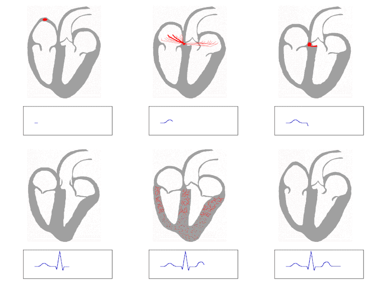
| Isolated Heart conduction system showing purkinje fibers | |
 | |
| The QRS complex is the large peak in the diagram at the bottom. |
For the nervous cells, see Purkinje cell
Purkinje fibers (Purkyne tissue or Subendocardial branches) are located in the inner ventricular walls of the heart, just beneath the endocardium. These fibers are specialized myocardial fibers that conduct an electrical stimulus or impulse that enables the heart to contract in a coordinated fashion.
Contents[hide] |
[edit] Histology
Purkinje fibers are a unique end-organ cardiac extension of the Autonomic Nervous System. Further histologic examination reveals that these fibers are split into left and right trees as well as atrial and ventricular contributions. The electrical origin of atrial Purkinje fibers arrives from the Sinoatrial Node. The following electrical origin of the ventricular Purkinje fibers arrives from the Atrioventricular Node.
Given no aberrant channels, the atrial and ventricular Purkinje trees are distinctly shielded from each other by collagen or the cardiac skeleton. The Purkinje fibers are uniquely dedicated to sympathetic electrical depolarization of the right and left atria and ventricles. The Purkinje fibers are further specialized to rapidly conduct impulses (numerous sodium ion channels and mitochondria, fewer myofibrils than the surrounding muscle tissue). Purkinje fibers take up stain differently than the surrounding muscle cells, and, on a slide, they often appear lighter and larger than their neighbours. They are binucleated.
[edit] Function
Heart rate is governed by many influences from the Autonomic Nervous System. The Purkinje Fibers do not have any known role in setting heart rate, but are influenced by Sympathetic discharge from the Sinoatrial node and thoracic Spinal Accessory Ganglia[dubious ].
During the ventricular contraction portion of the cardiac cycle, the Purkinje fibers carry the contraction impulse from both the left and right bundle branch to the myocardium of the ventricles. This causes the muscle tissue of the ventricles to contract, thus enabling a force to eject blood out of the heart; either to the Pulmonary circulation from the right ventricle or to the Systemic circulation from the left ventricle.
Atrial and ventricular discharge through the Purkinje trees is assigned on a standard Electrocardiogram as the P Wave and QRS complex, respectively.
Purkinje fibers also have the ability of automaticity[1], firing at a rate of 15-40 beats per minute if left to their own devices. In contrast, the SA node outside of parasympathetic control can fire a a rate of almost 100 beats per minute. - in short, they generate action potentials, but at a slower rate than sinoatrial node and other atrial ectopic pacemakers. Thus they serve as the last resort when other pacemakers fail. When a pukinje fiber does fire it is called a Premature ventricular contraction or PVC. Another name given is Ventricular escape.
[edit] Etymology
They were discovered in 1839 by Jan Evangelista Purkyně, who gave them his name.
[edit] See also
[edit] References
- ^ [biomed.engr.sc.edu/bme_lab/lab%20reports/36)%20ECG%20I.pdf ]
[edit] External links
- subendocardial+branches+of+atrioventricular+bundles at eMedicine Dictionary
- Organology at UC Davis Circulatory/heart/purkinje/purkinje1 - "Mammal heart, purkinje fibers (LM, Medium)"
- Anatomy Atlases - Microscopic Anatomy, plate 05.78
- MedEd at Loyola Histo/practical/cardio/hp8-21.html
- Histology at ucsd.edu
- Histology at nhmccd.edu
| ||||||||||||||||||||||||||||||||